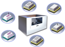
组织芯片仪
Automatically Relocates Tissues From Multiple Patients To One Slide. |
| |
 |
high-throughput Tissue Micorarray (Cat. No. : ATA-30)
Introduction
Several “high throughput methods” have been
introduced into research and routine laboratories during the past
decade.
These new techniques enhance the analysis and research of genomic
alterations and RNA or protein expression patterns in a relatively short
time, and promise to deliver clues to the diagnosis and treatment of
human cancer. Tissue microarray technology is one of these new tools. It
is a method of relocating multiple tissues from conventional
histological paraffin blocks so that tissues from multiple patients can
be seen on a same slide. The potential and the scientific value of
tissue microarray in modern research have been demonstrated in a
logarithmically increasing number of studies. Avans Biotech is proudly
to participate in this new era and ATA-30 will boost the tissue array
research programs significantly and automatically. |
|
|
Innovation Design |
New Innovation And Patented Technology
ATA is built on an innovative technology. It is a fully automatic
tissue arrayer with compact size that allows rapid and accurate
construction of low to high density tissue arrays. |
Easy To Use
Tissue array construction today is considered a tedious and
time-consuming task due to the intense concentration and complicates
operations. ATA-30 will boost this technology by providing fully
automatic operation. With the use of computer and software, all the
operation can be simplified by few mouse clicks. Computer file system
allows easy experiment saving and tracking. With the help of CCD vision
system, user will no longer require intense attention to the selection
and operation. This will completely revise your point of view and
approach to tissue array construction. Tissue array construction becomes
faster, easier and more precise. |
High Capacity
ATA-30 includes an minimum 18 blocks tray within the package in mode A system and additional tray is available.
You will be able to place any combination of recipient and donor blocks on the tray and define them in the software. |
Shorten Operation Time And Eliminate Manual Errors
You will be able to define where to collect and insert cores into donor or recipient block simply in the ATA-30 software.
This will streamline the operations, eliminate manual errors and shorten operation time.
ATA-30 is fully automatic that will increase the collection precision and guarantee high accuracy in histology type collected. |
CCD Vision System
ATA-30 is
integrated with CCD vision system that will allow you to operate all the
punch area selection directly from the screen easily. The CCD will also
record all the tissue blocks image and import into database. |
Integrated Software
The built-in software has two main functions: block database and tissue array designer.
| ‧ |
The database will allows you to manage and search the tissue
block with patient information easily and flexibly. Patients diagnosis
photo and tissue block photo is able to import and integrated into the
database. |
| ‧ |
The designer will allows you to generate any format of tissue
array from the entire patient database within a minute. The punch area
is selected directly on the screen by using mouse and the selection will
record inside the database for future avoidance. The depth and size of
the cores for each punch can be defined. |
|
 |
|
|
Method Comparison
|
Automated Arrayer |
Manual Arrayer |
Tracking |
Integrated software, export directly to a database |
Written notes |
Punch area selection |
Mark, edit and save punch coordinates using an digitally captured image from camera and software tools |
Visual selection while punching, using magnifying glass or a stereomicroscope as a guide and require intense concentration |
Pre-marking of punch areas |
CCD display pre-marked slide images side-by-side to the donor block image. |
Pathologist marks regions of interested to slides by hand before arraying |
Operation |
Computer controlled |
All movements performed manually by operator |
Punch speed |
Max 150~300 core per hour |
Max. 20~30 core per hour |
Block capacity |
Model A: max. 30 per tray
Model B: max. 42 per tray |
Max . 4 |
|
Technical specifications:
| Specifications |
| Power Input |
AC220V 300W |
| Dimensions |
W120 x D80 x H75 cm |
| Weight |
95kg |
| Platen |
30 blocks |
| Capacity |
30 blocks per tray with any combination of donor and recipient blocks. |
| Puncher Size |
0.6, 1.0, 1.5, 2.0 mm stainless needle sets |
| Speed |
150-300 cores transferred per hour |
| Operating temperature |
15-32℃ ambient |
| Operating humidity |
20-80% |
| Hardware |
| Working Range |
400 / 350 / 50 (mm) |
| Max. Loading |
XY 10kg / Z 5kg |
| Resolution |
0.01mm / Axis |
| Repeatability |
+/- 0.01 mm / Axis |
| Motor System |
Japan servo motor |
| Max. Speed |
XY-500 Z-250 (mm/sec) |
| Driving Method |
XYZ-Screwball axis |
| Loading Method |
Removable Tray with easy loading |
| Control Method |
PC - WinXP |
| Merging Method |
Low resolution CCD for stained slide and tissue block merging |
| Selection Method |
High resolution CCD for detail viewing and selection |
| Lighting Method |
Halogen ring light illumination |
| Interpolation Function |
3 axis |
| Software |
‧Enhanced software features
‧Tissue block database integrate with clinical and diagnosis data
‧Mark, edit and save punch coordinates using an on-screen display and software tools
‧Tissue punch coordinates history
‧Easy array programming with punch size selection and core annotation
‧Save workflow in file for repeat arraying
‧Advance and easy punch area selection
‧Export block image and data |
| Optional |
‧Additional tissue block tray
‧Constant temperature tray and heating device
‧2.0 and 2.5 mm stainless needle sets
‧Barcode system integration |
| Warrenty |
1 year warranty |
| Package |
Automatic Tissue Arrayer |
| 3 sets of punch needles ( 0.6mm , 1.0mm , 1.5mm ) |
|

